
Why biomimetic blockcopolymer membranes?
Transmembrane proteins fulfill plenty tasks in living organisms. For example, the exchange of substances between the cells and their surrounding is controlled and regulated or chemical energy in form of ATP is regenerated in the cells by transmembrane proteins. Some membrane proteins possess high selectivities for ions or small molecules like H2O.
Such selectivities triggered interest for the membrane proteins also in the field of technical processes. For example in nanofiltration, a high selectivity and salt rejection is a crucial point that could be fulfilled with membrane proteins. However, natural lipid bilayers, are not stable enough in technical processes. One alternative for lipid bilayers are amphiphilic blockcopolymers. Under certain conditions – concentration, molecular weight, block ratio – they can form biomimetic bilayered structures in water.
If the successful combination of functional transmembrane proteins and biomimetic blockcopolymer bilayers is achieved, for example, new filtration processes are possible.
Therefore, we synthesize amphiphilic blockcopolymers using anionic polymerization methods and study the self-assembly of these materials in water towards the formation of biomimetic membranes.
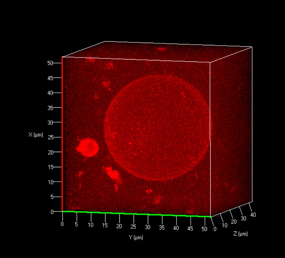
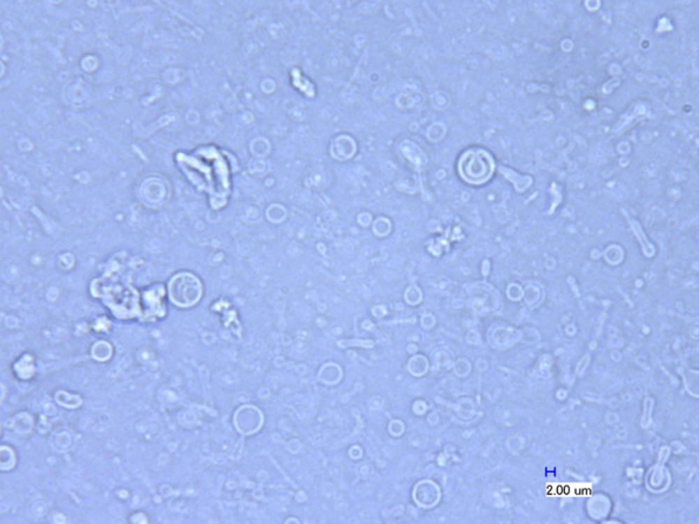
Depending on blockcopolymer composition, different self-assembled structures can be obtained. Either giant unilamellar vesicles up to 80 µm in diameter (Figure 1) or manifold smaller unilamellar structures can be formed (Figure 2).

Membrane characterization
The characterization of biomimetic blockcopolymer membranes focused on the detection of functional incorporated membrane proteins represents another main part of this project.
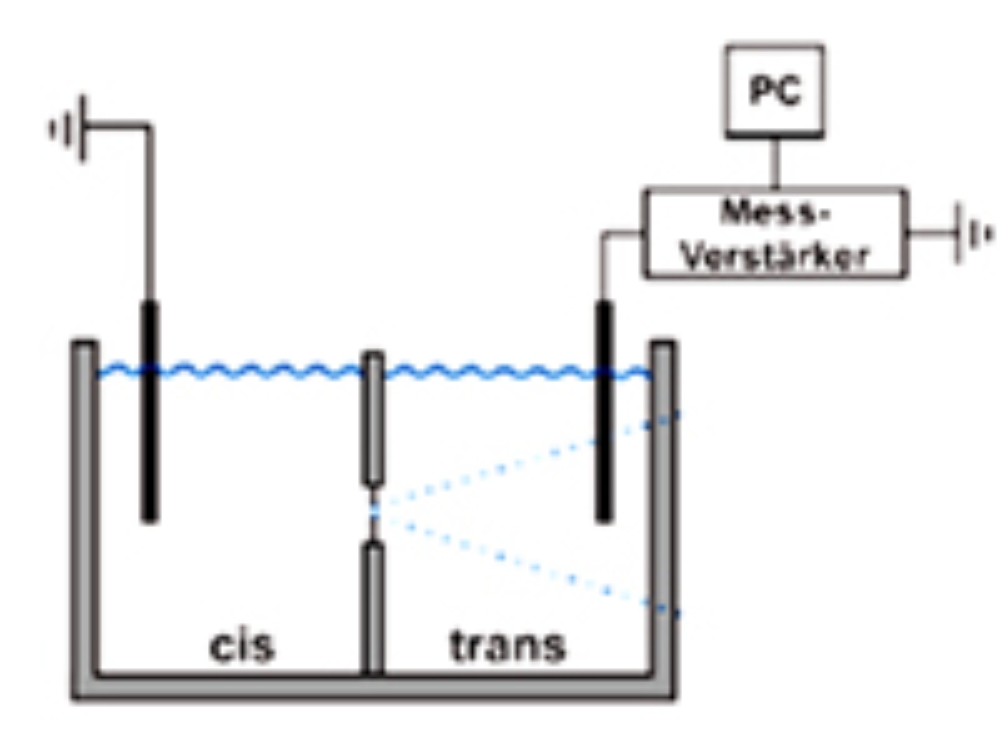
Therefore, planar blockcopolymer membranes are characterized with electrochemical methods like impedance spectroscopy and membrane thicknesses and resistances extracted from the obtained data (Figure 3).
Membranes currently under investigation have thicknesses between 8 to 15 nm, depending on molecular weight.
Reconstitution of transmembrane proteins
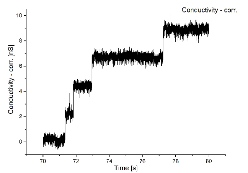
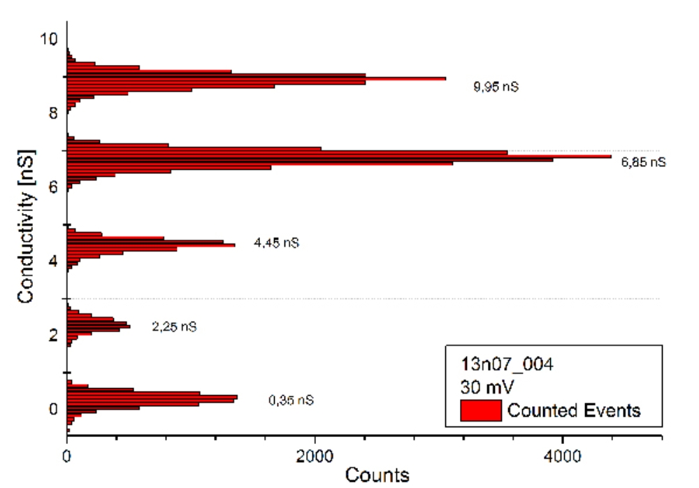
To proof the functional reconstitution of membrane proteins into biomimetic blockcopolymer membranes, the porin OmpF from E. coli is used. Funcional reconstitution is not mandatory, since the blockcopolymer membranes show higher thicknesses as natural lipid membranes.
Nevertheless, the functional reconstitution of OmpF was successful for the first time using the studied amphiphilic blockcopolymer.
Thereby, the conductivity across the membrane shows characteristic jumps (Figure 4)
Funding
- Kekulé-Felloship of the Chemical Industry Fund
- Fraunhofer „Cell-free bioproduction“
- Federal Ministry of Education and Research Germany (BMBF)
Partners
This research is performed in close collaboration with the following partners:
- Dr. Thomas Schiestel and Dr. Michaela Müller, Fraunhofer IGB Department of Interfacial Engineering and Materials Science
- Dr.-Ing. Christina Weber, Fraunhofer IGB Department of Molecular Biotechnology
- Prof. Dr. Stephan Nußberger, Biophysics Department, Biological Institute, University Stuttgart
- Dr. Eric Nebling, Fraunhofer ISIT, Department of Biotechnical Microsystems
Duration:
- 2012 bis 2015

Alexander Southan
Dr.Coordination of Chemical-Physical Interfaces // Head of NanoBioMater